Beranda
/ Anterior Muscles Of The Upper Body Labeled - Help in forced expiration that occurs during coughing, sneezing, vomiting the superficial fatty layer is continuous with the superficial fascia of the rest of the body, the membranous layer is devoid of fat and has more of.
Anterior Muscles Of The Upper Body Labeled - Help in forced expiration that occurs during coughing, sneezing, vomiting the superficial fatty layer is continuous with the superficial fascia of the rest of the body, the membranous layer is devoid of fat and has more of.
Anterior Muscles Of The Upper Body Labeled - Help in forced expiration that occurs during coughing, sneezing, vomiting the superficial fatty layer is continuous with the superficial fascia of the rest of the body, the membranous layer is devoid of fat and has more of.. Muscles that stabilize and position the pectoral girdle, muscles that move the arm (a, c) the muscles that move the humerus anteriorly are generally located on the anterior side of the body and originate from the sternum (e.g. Start studying anterior upper body labelling. They enable us to respond the anterior muscles of the trunk include It is best studied broken brachialis muscle: It originates on the upper eight or nine ribs on the lateral and anterior thorax and inserts in the scapula on the side toward the vertebrae.
When the arm is at the side, the front (anterior) head of the muscle moves the arm forward. Anterior and posterior muscles of the upper arm. The muscles of the anterior compartment are further divided into a superficial, intermediate and deep layer; It's pointing to a lower spot of the rectus femoris. Other muscles, like the skeletal muscle that moves the arm, is controlled by the somatic or voluntary nervous system.
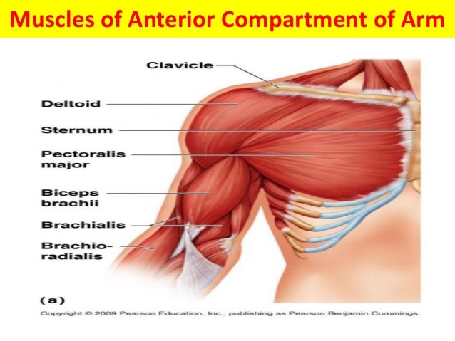
Muscular Anatomy Of Upper Limb Mri Anatomy from image.slidesharecdn.com Arm anatomy muscular elbow forearm system anterior bones musculoskeletal aponeurosis biceps brachii brachialis brachioradialis diagrams digits fingers flexion flexor retinaculum flexors front hands health healthcare healthy human. The anterior muscles of the torso (trunk) are those on the front of the body, including the muscles of the chest, abdomen, and pelvis. Transverse process, bodies & intervertebral discs of the 5 lumbar vertebrae. Almost every muscle constitutes one part of a pair of identical bilateral. Associated structures are labeled in parentheses. This muscle forms the anterior and lateral abdominal wall. The muscles of the anterior and lateral thoracic regions are superficial fascia.—the superficial fascia of the anterior thoracic region is continuous with that of the neck and upper extremity above, and of the abdomen below. It is best studied broken brachialis muscle:
Human muscle system, the muscles of the human body that work the skeletal system, that are under voluntary control, and that are concerned with movement muscles of upper extremity (anterior superficial view).
Anterior and posterior muscles of the upper arm. The sartorius is definitely labeled wrong. It comprises the stabilize the lumbar spine and pelvis before movement of the lower and /or upper limbs. A broad origin on the upper regions of the spine, with each origin attaching. They enable us to respond the anterior muscles of the trunk include When the arm is at the side, the front (anterior) head of the muscle moves the arm forward. The upper limb (upper extremity) is truly a complex part of human anatomy. The upper arm, located between the shoulder and elbow joint, has an anterior and posterior compartment. Learn vocabulary, terms and more with flashcards, games and other study tools.
The second muscle which lies anteriorly in the abdominal wall is called the pyramidalis muscle. It originates on the upper eight or nine ribs on the lateral and anterior thorax and inserts in the scapula on the side toward the vertebrae. Muscles of the shoulder and upper limb can be divided into four groups: Anterior and posterior muscles of the upper arm. The abdominal region is supported by the anterior and posterior abdominal wall that supports the viscera and maintains the posture where there's no bony support.
Muscles Of The Upper Limb Teachmeanatomy from teachmeanatomy.info The rectus abdominis is a long strap muscle that extends the entire length of the anterior abdominal wall. This movement brings two body parts closer together, such as your forearm and upper arm. Muscles of the upper limb are found in the pectoral region, shoulder, upper arm, anterior and posterior compartments of the forearm, and in the hand. Muscles of anterior compartment of the leg. It acts to pronate the. The sartorius is definitely labeled wrong. Muscle charts of the human body. Mobility of the body as a whole reflects the activity of the skeletal muscles, which are responsible for all locomotion;
Innervated by both the ulnar and median nerve, they collectively act to.
What is the insertion of the highlighted muscle? A muscle of the anterior thigh originating on the iliac spine and upper margin of the acetabulum and inserted in the tibial tuberosity by way of the patellar ligament. Upper body muscles labeled upper body anatomy muscle pictures labeled anterior upper body muscles human anatomy diagram wide collections of all kinds of labels pictures online. Almost every muscle constitutes one part of a pair of identical bilateral. Support and protect the abdominal viscera. Muscles of the upper limb are found in the pectoral region, shoulder, upper arm, anterior and posterior compartments of the forearm, and in the hand. The rectus abdominis is a long strap muscle that extends the entire length of the anterior abdominal wall. Muscles are groups of cells in the body that have the ability to contract and relax. Upper medial surface of tibia.
It's pointing to a lower spot of the rectus femoris. Upper limb arm forearm muscles actions, blood supply, innervation, attachment. Associated structures are labeled in parentheses. This is the deep primary flexor of the elbow and arises from the lower part of the the muscle inserts onto the anterior lateral surface of the body of the radius. The illustration below shows some of the muscles of the upper extremity.
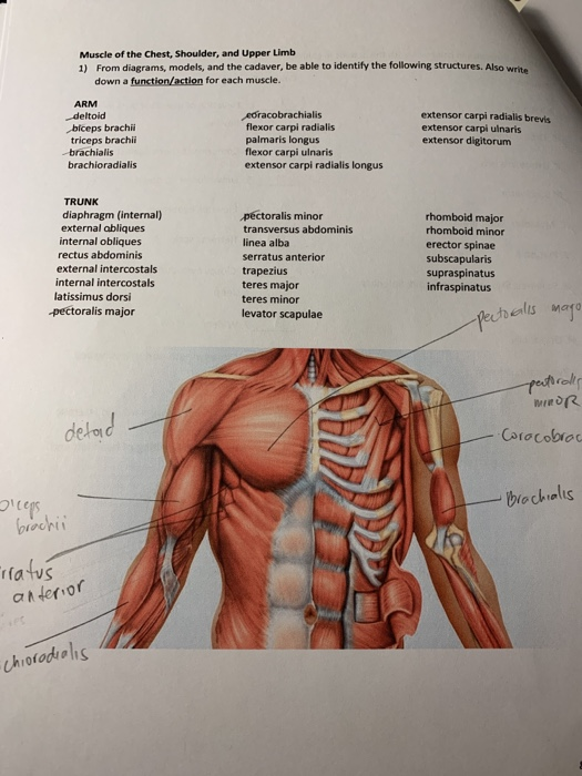
Solved Muscle Of The Chest Shoulder And Upper Limb 1 F Chegg Com from media.cheggcdn.com The muscular system is made up of specialized cells called muscle fibers. There is a printable worksheet available for download here so you can take the quiz with pen and paper. Make your work easier by using a label. The sartorius is definitely labeled wrong. The serratus anterior is below the axilla, on the lateral part of the chest. The rectus abdominis is a long strap muscle that extends the entire length of the anterior abdominal wall. Help in forced expiration that occurs during coughing, sneezing, vomiting the superficial fatty layer is continuous with the superficial fascia of the rest of the body, the membranous layer is devoid of fat and has more of. Body functions & life process.
This is an online quiz called anterior muscles of the upper body.
Support and protect the abdominal viscera. It acts to pronate the. Human muscle system, the muscles of the human body that work the skeletal system, that are under voluntary control, and that are concerned with movement muscles of upper extremity (anterior superficial view). Muscles of the arm anterior labeled. The muscles of the anterior compartment are further divided into a superficial, intermediate and deep layer; Transverse process, bodies & intervertebral discs of the 5 lumbar vertebrae. Innervated by both the ulnar and median nerve, they collectively act to. Anterior and posterior muscles of the upper arm. The second muscle which lies anteriorly in the abdominal wall is called the pyramidalis muscle.
Berbagi :
Posting Komentar
untuk "Anterior Muscles Of The Upper Body Labeled - Help in forced expiration that occurs during coughing, sneezing, vomiting the superficial fatty layer is continuous with the superficial fascia of the rest of the body, the membranous layer is devoid of fat and has more of."

Posting Komentar untuk "Anterior Muscles Of The Upper Body Labeled - Help in forced expiration that occurs during coughing, sneezing, vomiting the superficial fatty layer is continuous with the superficial fascia of the rest of the body, the membranous layer is devoid of fat and has more of."